normal range echocardiography normal values pdf
AIMS Availability of normative reference values for cardiac. Several accredited echocardiography laboratories of the European Association of Cardiovascular Imaging EACVI7 The NORRE study aims to prospectively establish a set of normal echocardio-graphic values in a large cohort of healthy individuals over a wide range of ages.

Pdf Normal Reference Intervals For Cardiac Dimensions And Function For Use In Echocardiographic Practice A Guideline From The British Society Of Echocardiography Semantic Scholar
Therefore in individuals aged 20 years EF in the range of 53 to 73 should be classified as normal.
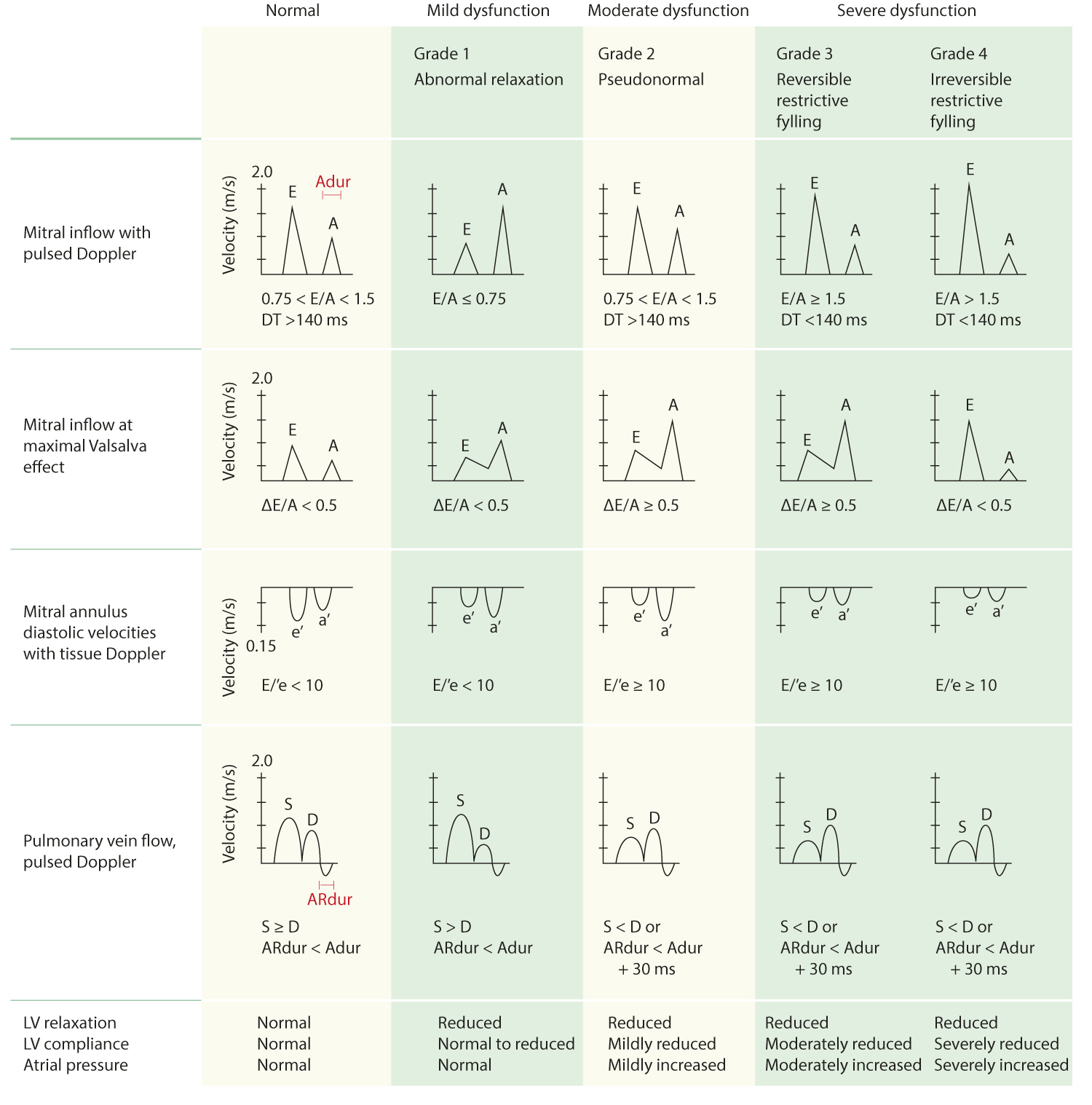
. A TAPSE cutoff value 17 mm yielded high specificity though low sensitivity to. Normal reference ranges for cardiac chambers size obtained in a large group of healthy volunteers accounting for gender and age highlight the need for body size normalization that should be performed together with age-and gender-specific assessment for the most echocardiographic parameters. Impaired relaxation EA -.
A Z score of 2 or 2 corresponds to the 95th percentile ie 2 standard deviations above or below the mean Nomograms and Z scores. Aggarwal S Pettersen MD Gurckzynski J et al. Z score is 0.
ECHOCARDIOGRAPHY WASE NORMAL VALUES STUDY As proposed in the WASE Normal Values Study many of these lim-itations will be addressed by performing a head-to-head comparison in which all technical differences in data acquisition between regions will be minimized and standardized by following strict ASEEACVI. The analyses were restricted to healthy participants. Aortic Annulus Size 18-23 cm Mitral Annulus Size 30-35 cm Aortic VTI 18-25 cm Mitral VTI 10-13 cm.
Normal Echo Values - Discover Echo. However due to the lack of consistency in current echocardiographic reference values their use for clinical decision-making remains questionableAimsThe aim of the Normal Reference Ranges for. Normal Echocardiographic Values for Cardiovascular Structures 891 2SD 2SD Mean 00 10 20 30 Aortic Sinotubular Junction cm 40 00 05 10 15 20 25 Body Surface Area m2 Figure A22 Inner-edge to inner-edge mid-systolic aortic sinotubular junction diameter versus body surface area.
2SD 2SD Mean Body Surface Area m2 0 1 2 3 4. Development of reference limits1419 The Normal Reference Ranges for Echocardiography Study NORRE Study aims to pro-spectively provide a set of normal values in a large cohort of healthy individuals over a wide range of ages 2575 years old. Normal Echo Sample ID.
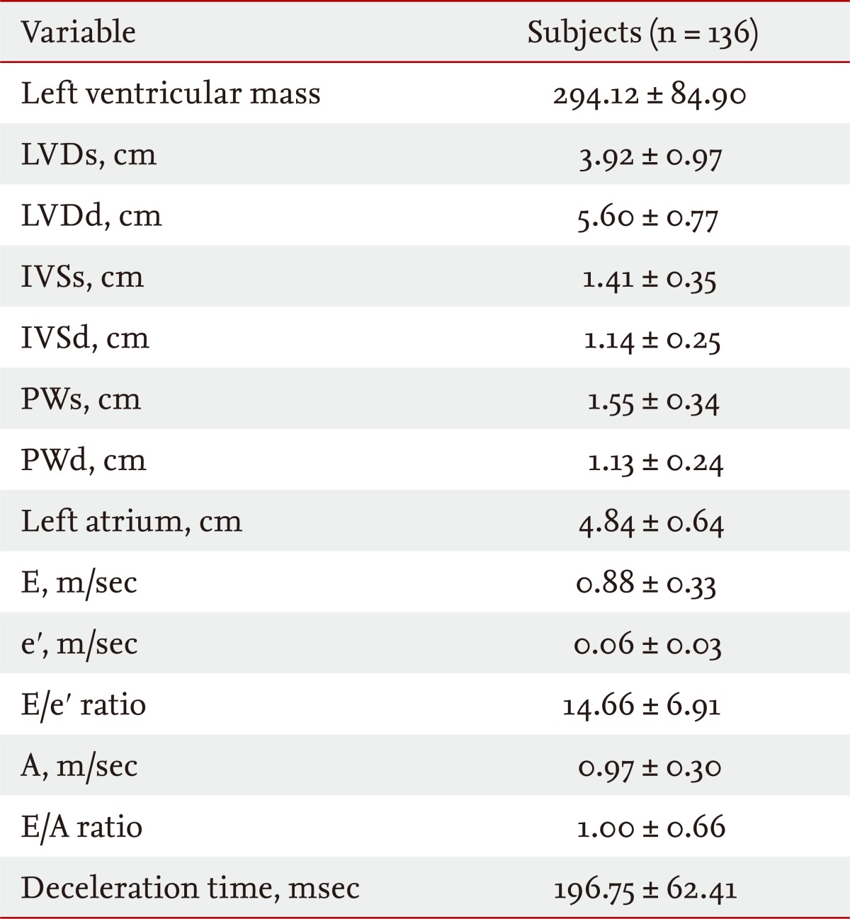
Recently both the 2D chamber size and Doppler. The left ventricle is normal in size with normal function. Three-dimensional Table 2 Normal values for 2D echocardiographic parameters of LV size and function according to gender.
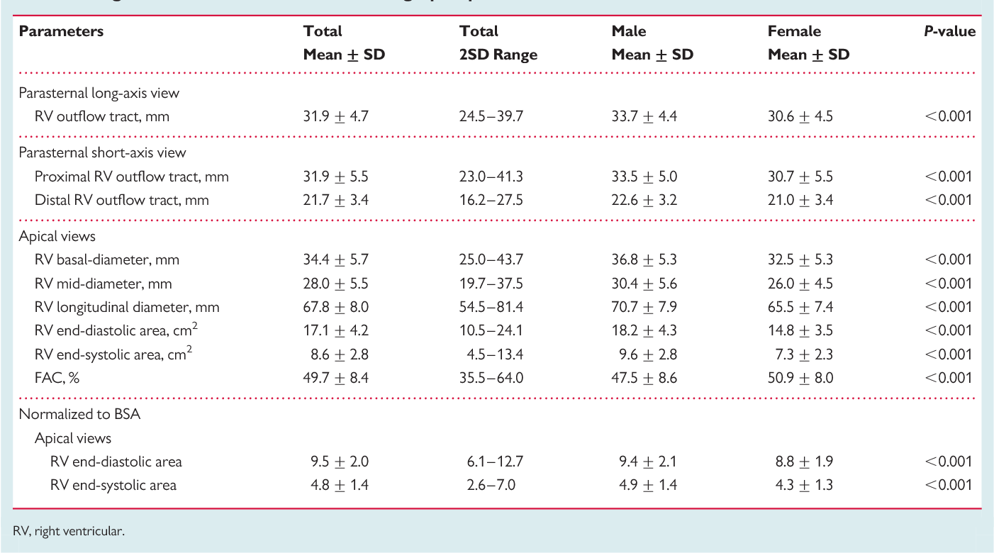
New upper reference limits for RV outflow tract dimensions RV body and the right atrium. An Update from the American Society of Echocardiography and the European Association of Cardiovascular Imaging 2015 JASE 2015 Jan2811-39e14. Method to measure the distance of systolic excursion of the RV annular segment along its longitudinal plane from a standard apical 4- chamber window.
Normal EF was 63 6 5 using the biplane method of disks. LV Dimensions Volumes Mass Normal Mild Moderate Severe Normal Mild Moderate Severe LVIDdiastolemm 3756 5761 6265 65 3551 5255 5659 59 LVIDsystolemm 2241 4245 4650 50 2037 3842 4346 46. Echocardiographic measurements were described by mean standard deviation 95 percentile and 95 confidence limits.
Separate reference intervals for males and females. Echocardiographys Guidelines and Standards Committee and the Chamber Quantification Writing Group developed in con-junction with the European Association of Echocardiography a branch of the European Society of Cardiology. The base of the RV free wall provides one of the most visibly obvious movements on normal echocardiography.
Thus to normalize in pediatric echocardiography we use nomograms Normalized data are expressed as z score ie. To trust in z scores. The left ventricular wall mo-tion is normal.
Introduction of indexed values to allow for body habitus. Normal EA 10-20 2. Normal Area 25-45 cm2 Mild Stenosis 10-25 cm2.
- Recommendations for the Evaluation of Left Ventricular Diastolic Function by Echocardiography. A company is evaluating if E-Echo will continue past Dec 31 2022. Routine abnormal stress test.
A total of 100 healthy participants with several characteristics similar to those from the original population had a complete and reliable echocardiographic. A z score 2 indicate dilatation while a z score. Changes in the echocardiographic assessment of the right heart.
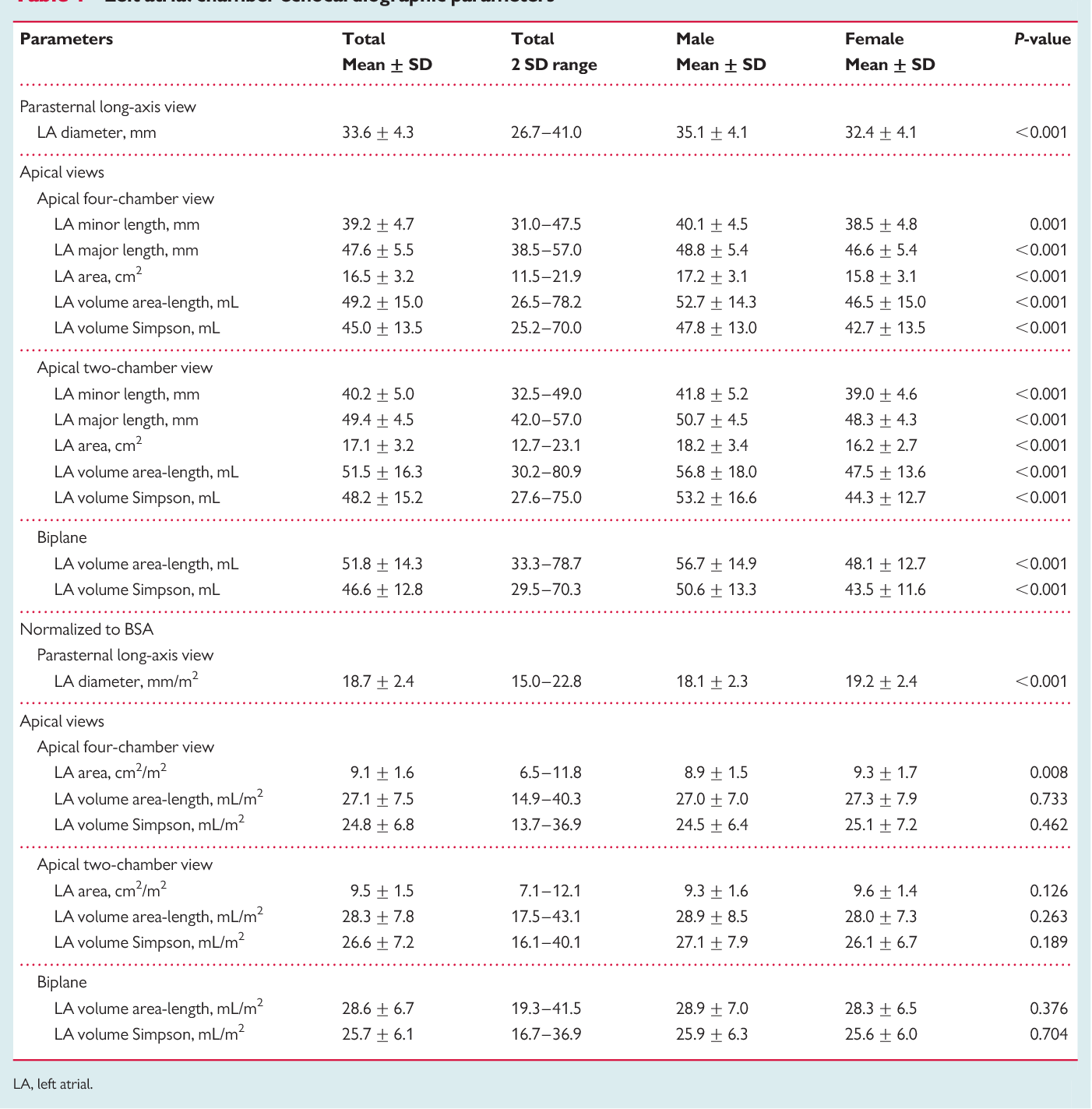
J Am Soc Echo-cardiogr 200518144063. The ejection fraction is 65. A new borderline LA volume range of 34-38mlm 2.
An Update from the American Society of Echocardiography and the. Restrictive filling EA - 20.
Pdf Echocardiographic Reference Ranges For Normal Cardiac Chamber Size Results From The Norre Study Semantic Scholar

Pdf Normal Echocardiographic Measurements In Indian Adults How Different Are We From The Western Populations A Pilot Study

Pdf Normal Echocardiographic Measurements In Indian Adults How Different Are We From The Western Populations A Pilot Study
Reference Normal Values For Echocardiography Ecg Echo

Pdf Echocardiographic Reference Ranges For Normal Cardiac Doppler Data Results From The Norre Study

Pdf Echocardiographic Reference Ranges For Normal Cardiac Chamber Size Results From The Norre Study Semantic Scholar

Echocardiography Normal Values Techmed

Pdf Echocardiographic Reference Ranges For Normal Cardiac Doppler Data Results From The Norre Study

Pdf Echocardiographic Reference Ranges For Normal Cardiac Doppler Data Results From The Norre Study Semantic Scholar

Pdf Echocardiographic Reference Ranges For Normal Cardiac Doppler Data Results From The Norre Study

Pdf Echocardiographic Reference Ranges For Normal Cardiac Chamber Size Results From The Norre Study Semantic Scholar
Pdf Echocardiographic Reference Ranges For Normal Cardiac Chamber Size Results From The Norre Study Semantic Scholar

Pin By Jeremy Jaramillo On Medical Echocardiogram Vascular Ultrasound Cardiac Nursing

Table 2 From Percentile Curves Of Normal Values Of Echocardiographic Measurements In Normal Children From The Central Southern Region Of The State Of Sao Paulo Brazil Semantic Scholar

Pdf Echocardiographic Reference Ranges For Normal Cardiac Doppler Data Results From The Norre Study

E And E Wave Parameters Download Table

Pdf Echocardiographic Reference Ranges For Normal Cardiac Chamber Size Results From The Norre Study Semantic Scholar

Pdf Echocardiographic Reference Ranges For Normal Cardiac Chamber Size Results From The Norre Study Semantic Scholar
Tissue Doppler Derived E E Ratio As A Parameter For Assessing Diastolic Heart Failure And As A Predictor Of Mortality In Patients With Chronic Kidney Disease